
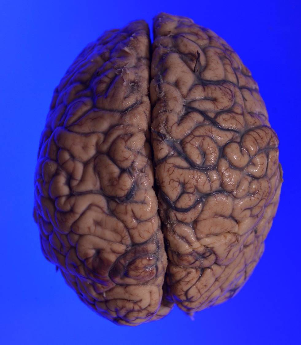


Superior and basal view of adult human brain
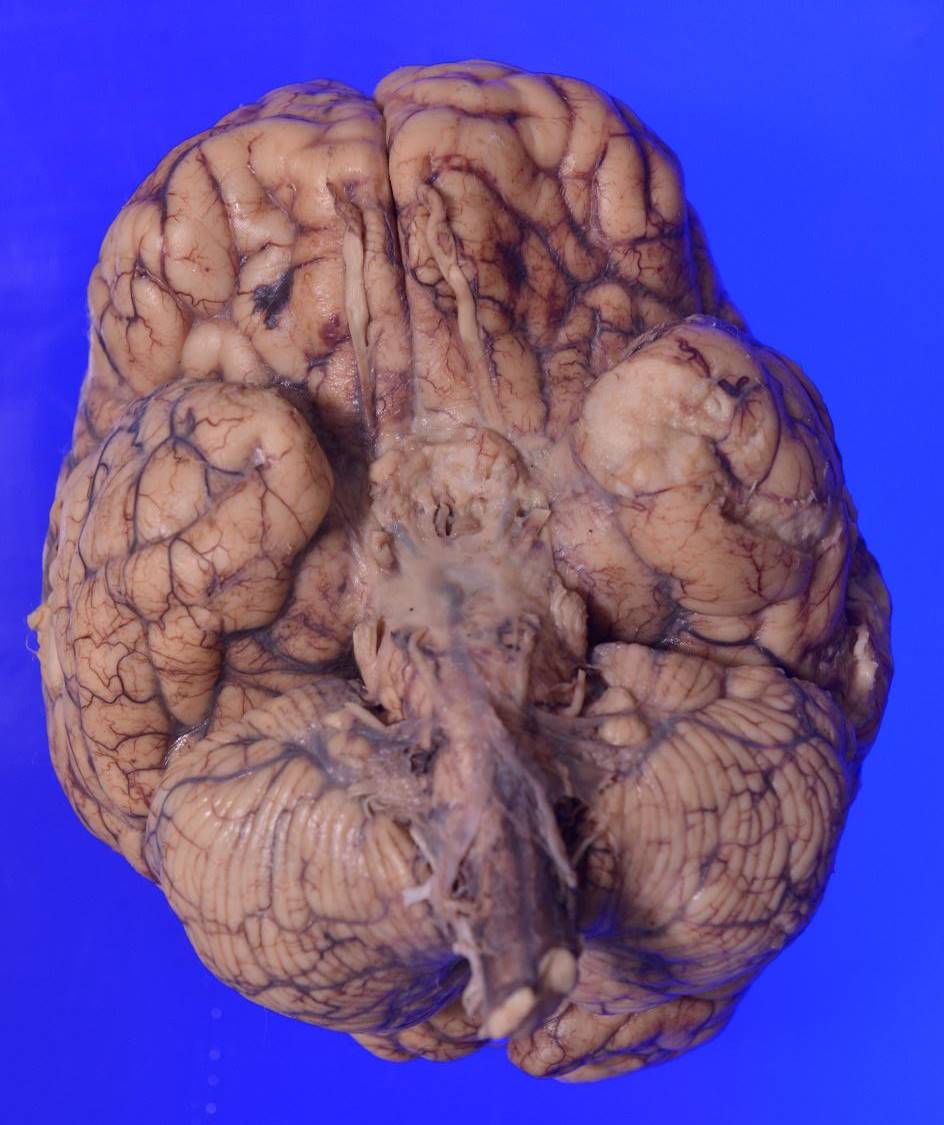
Superior and basal view of adult human brain
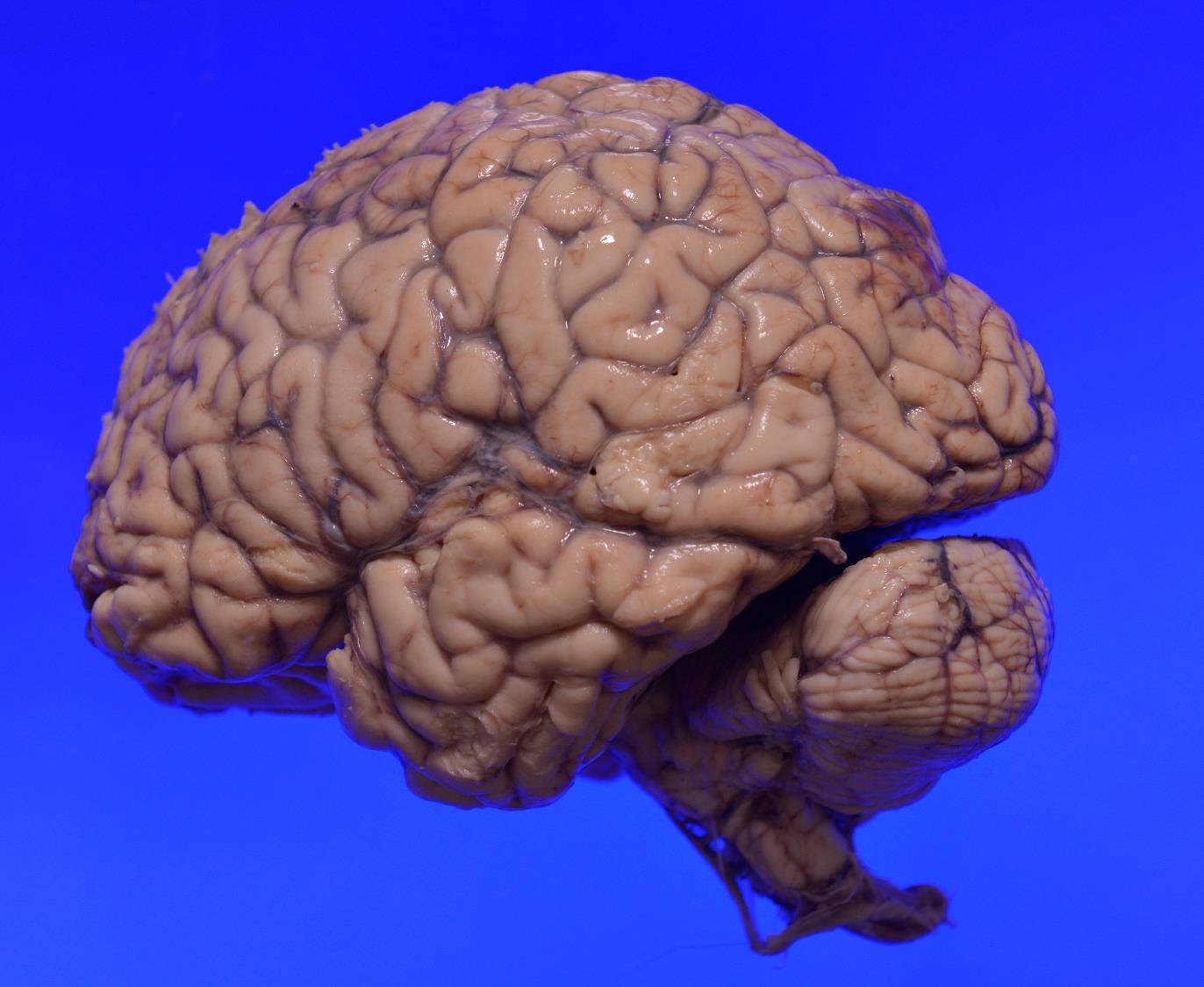
Lateral view of adult human brain
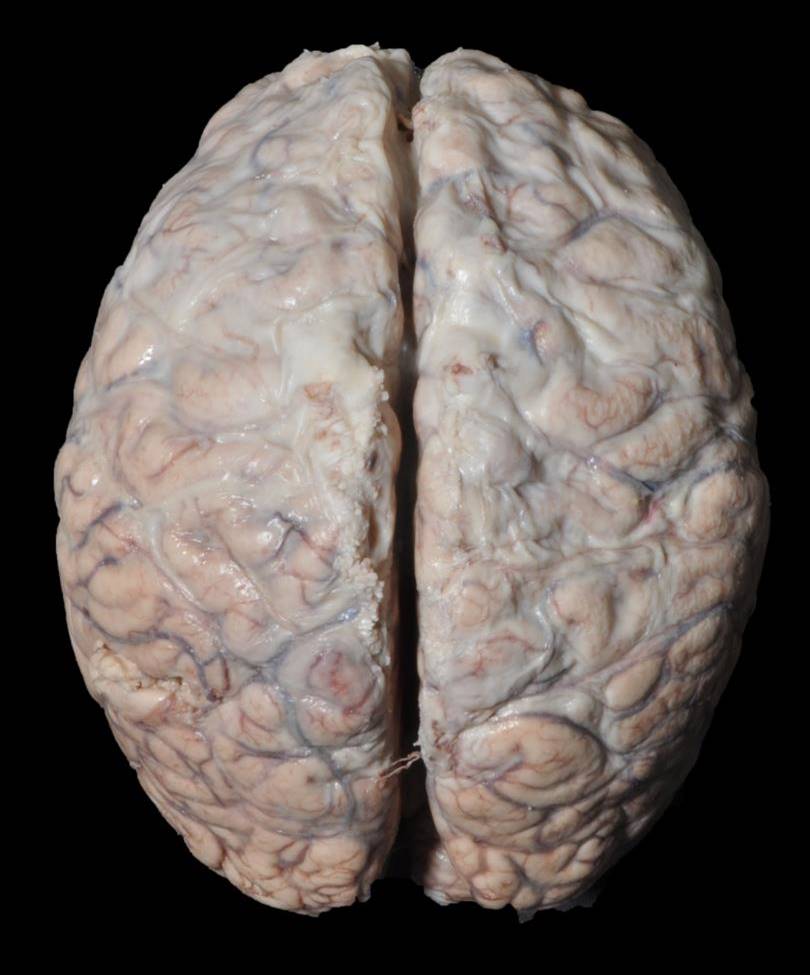
Superior and basal view of adult human brain
A case of pyogenic meningitis showing exudates on thecerebral convexities
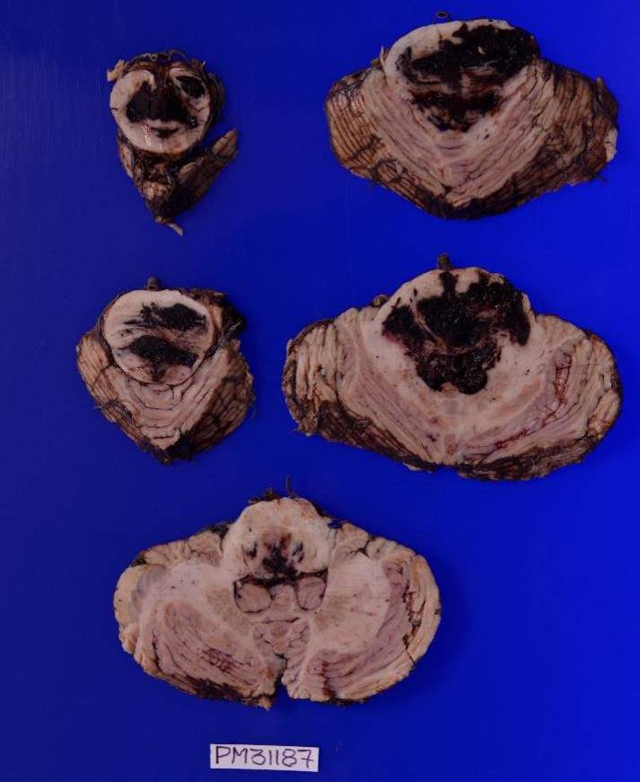
Axial section from brainstem showing hypertensive hemorrhage in the pons, medulla, with extension into the fourth ventricle.
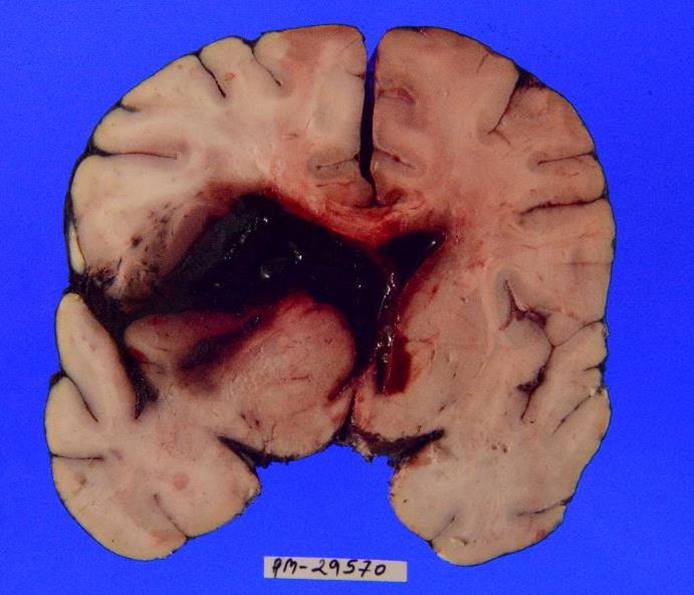
Coronal slice of brain showing large hypertensive intracerebral hemorrhage in left basal ganglia region with intraventricular extension and midline shift towards right side
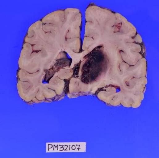
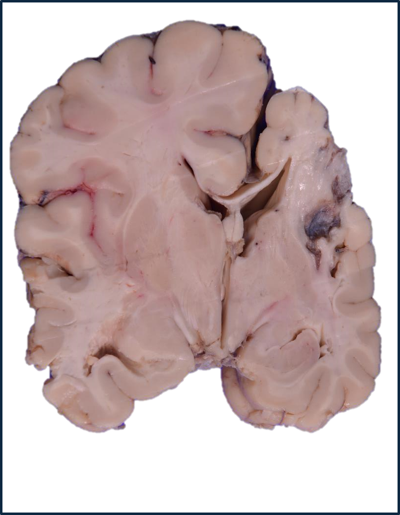
Coronal slice of brain showing hypertensive hemorrhage in right thalamus and internal capsule
Coronal slice of brain showing multiple bilateral embolic infarcts
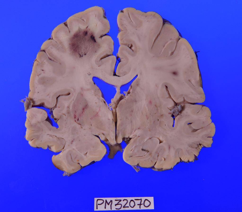
Coronal slice of brain showing an embolic infarct in the left frontal lobe
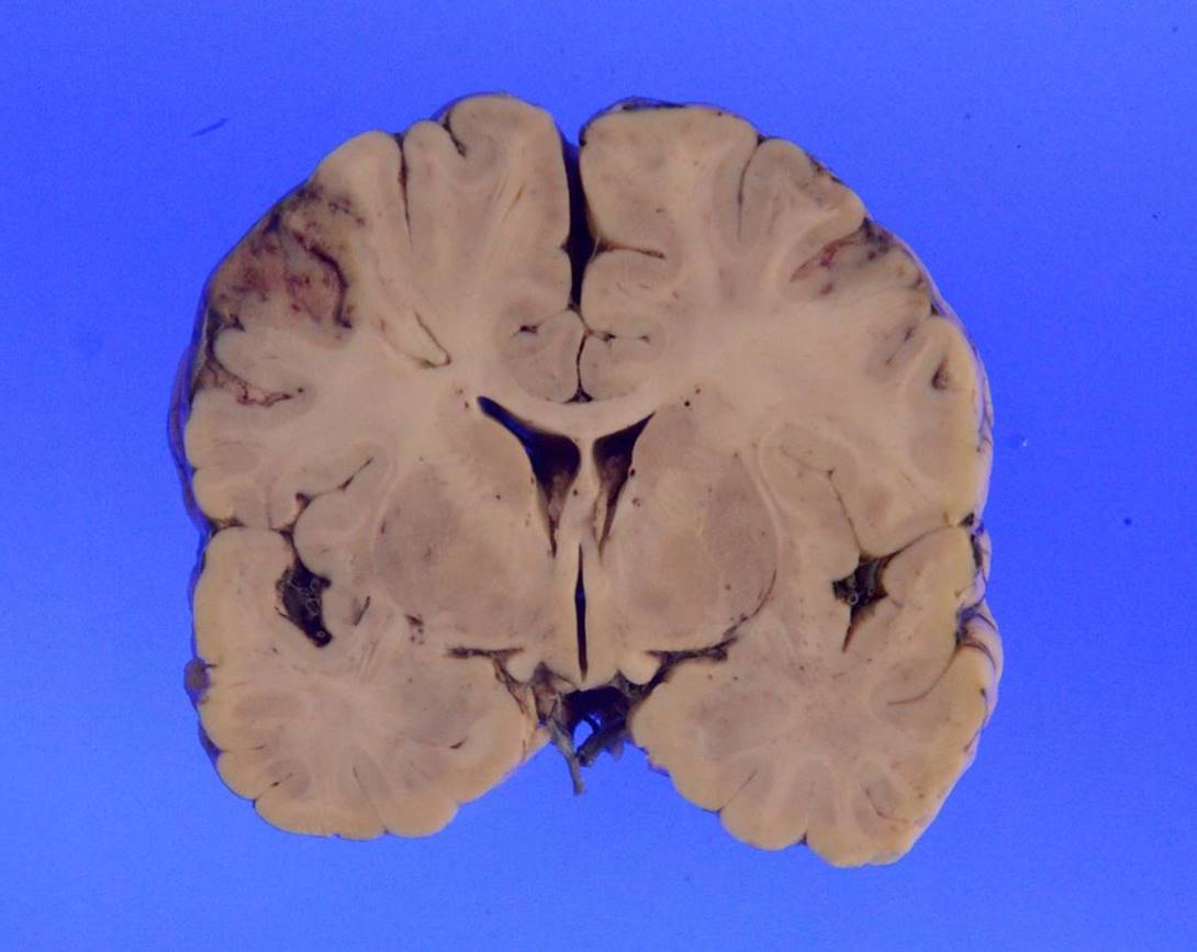
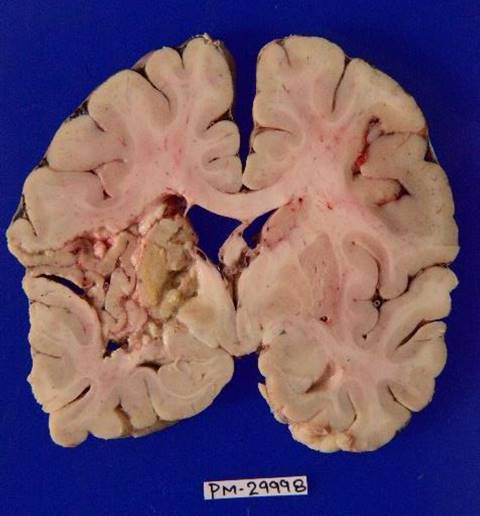
Coronal slice of brain showing an old infarct in the right middle cerebral artery territory
Coronal slice of brain showing an old cystic infarct in the left basal ganglia region
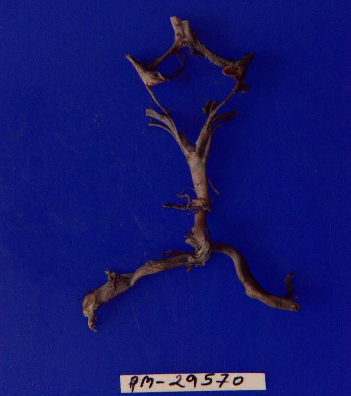
Circle of Willis in a case of ischaemic stroke showing atheromatous changes
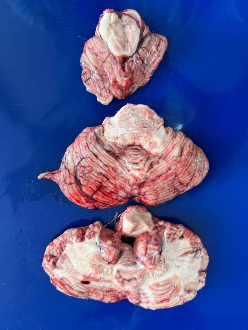
Axial sectioning of brainstem in fresh state
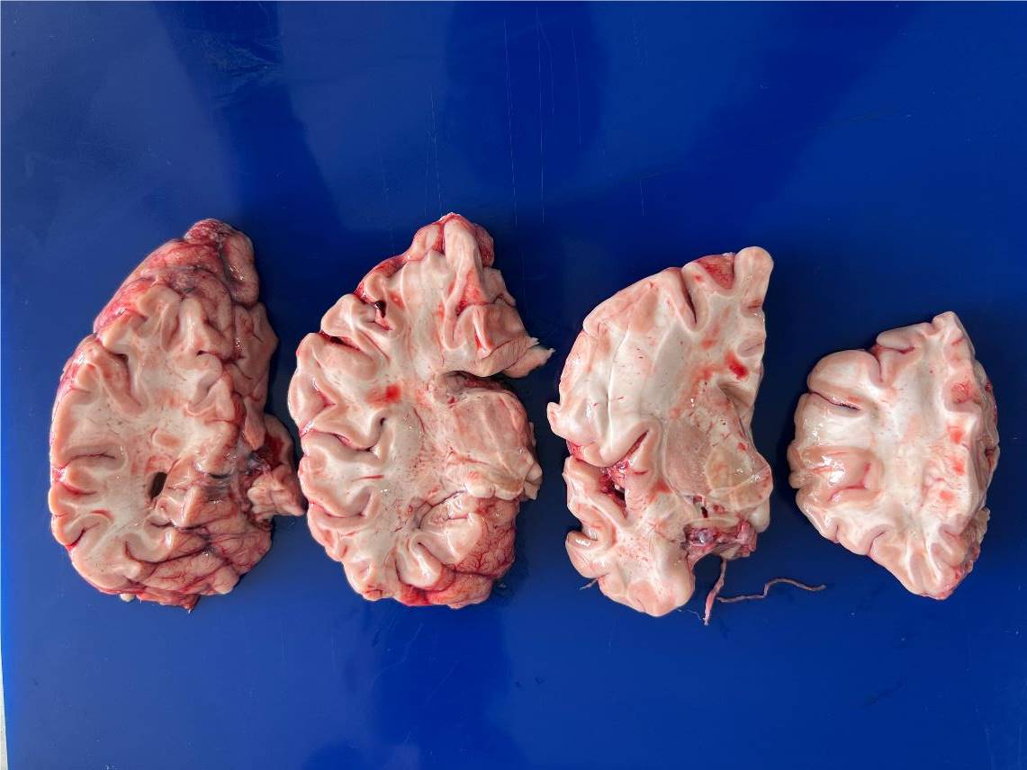
Coronal slice of brain in fresh state
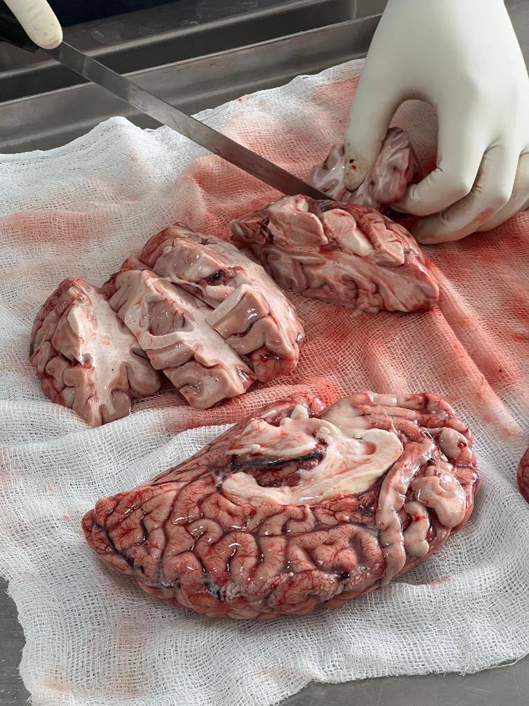
Brain is being cut in the coronal plane in the fresh state
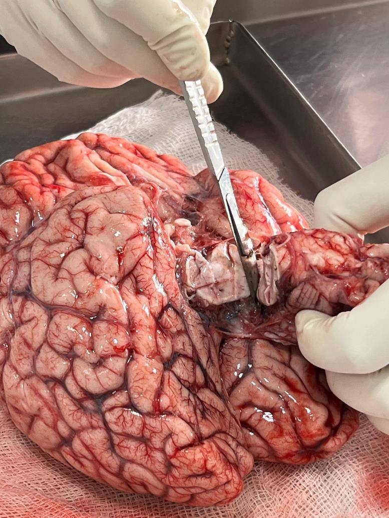
Brainstem is being separated from the brain
A case of tuberculous meningitis showing thick basal exudates covering the interpeduncular fossa